Preview image. Unlock full-res
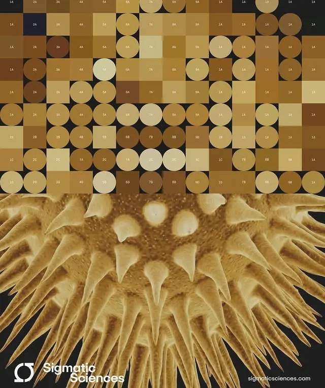
Scientific illustration splitting a geometric grid above with a magnified diatom photomicrograph below, bridging abstract and biological visualization.
Summary
A two-panel composition combining a labeled grid taxonomy of diatom specimens above with a detailed photomicrograph of actual diatom fiber structure below, bridging diagrammatic taxonomy and microscopic reality.
Visual description
The upper half organizes tan and cream colored circles and ovals on a dark ground in a structured grid, each labeled with abbreviations (1A, 2A, etc.), resembling a reference catalog or specimen chart. The lower half transitions into a photographic-quality magnified view of actual diatom fibers in warm gold and tan tones, revealing intricate radial striations and organic geometry. The Sigmatic Sciences wordmark appears in white sans-serif at the lower right. The color palette is uniformly warm and earthy across both sections, with contrast coming from the dark ground and the bright cream highlights on the specimens.
Key takeaway
The visual hierarchy of abstraction to detail, moving from symbolic representation to photographic evidence; the use of a consistent warm monochromatic palette to unify disparate visual languages; the contrast between regular grid spacing and organic texture as a visual device for showing classification versus observation.
Reuse notes
Excellent for educational, scientific, or medical materials where credibility and precision matter. Use when bridging conceptual frameworks with empirical evidence. The warm earth tones lend authority without coldness; ideal for life sciences, biology, materials science, or medical research presentations. Requires high-quality photography of actual specimens to work at this level of detail.
More like this






Keep what you find
A free account saves references to boards and can send you a weekly email of picks based on what you save.
To see every result, go Pro.